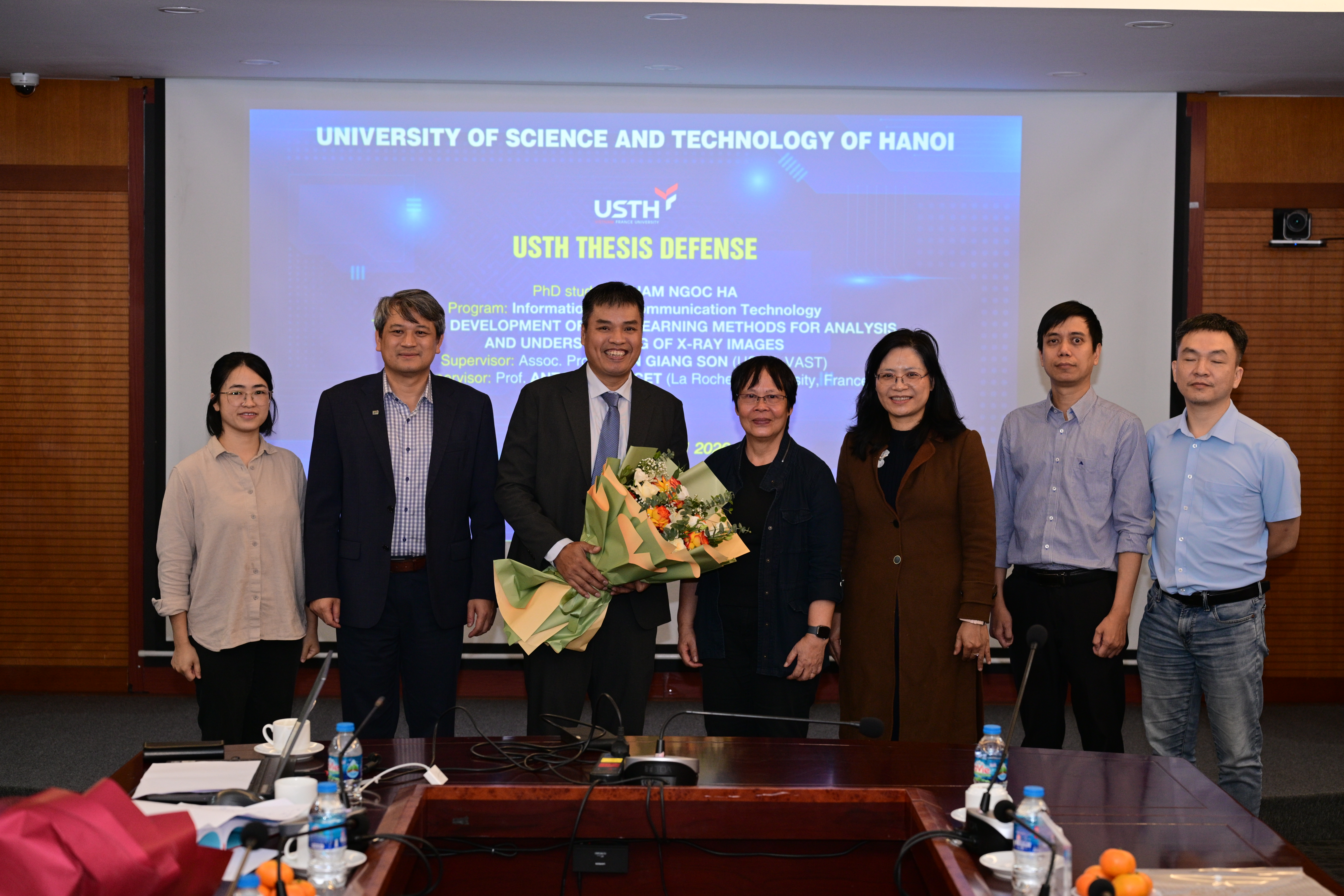
On November 11, 2026, the University of Science and Technology of Hanoi (USTH) held the university-level doctoral thesis defense of PhD candidate Pham Ngoc Ha in Information and Communication Technology. The thesis is titled “Development of deep learning methods for analysis and understanding of X-ray images.”
The research was supervised by Assoc. Prof. Tran Giang Son, Director of the Department of Information and Communication Technology, and co-supervised by Prof. Antoine Doucet, Co-Director of the Department and Vice-President for the European University EU-CONEXUS at La Rochelle University.

A defense following academic procedures
The thesis examination committee consisted of seven members and was chaired by Assoc. Prof. Luong Chi Mai from the Institute of Information Technology, Vietnam Academy of Science and Technology.
Under the leadership of the committee chair, the defense followed the required procedures. These included a review of Pham Ngoc Ha’s academic background, doctoral training, research results, and scientific publications during the PhD program.
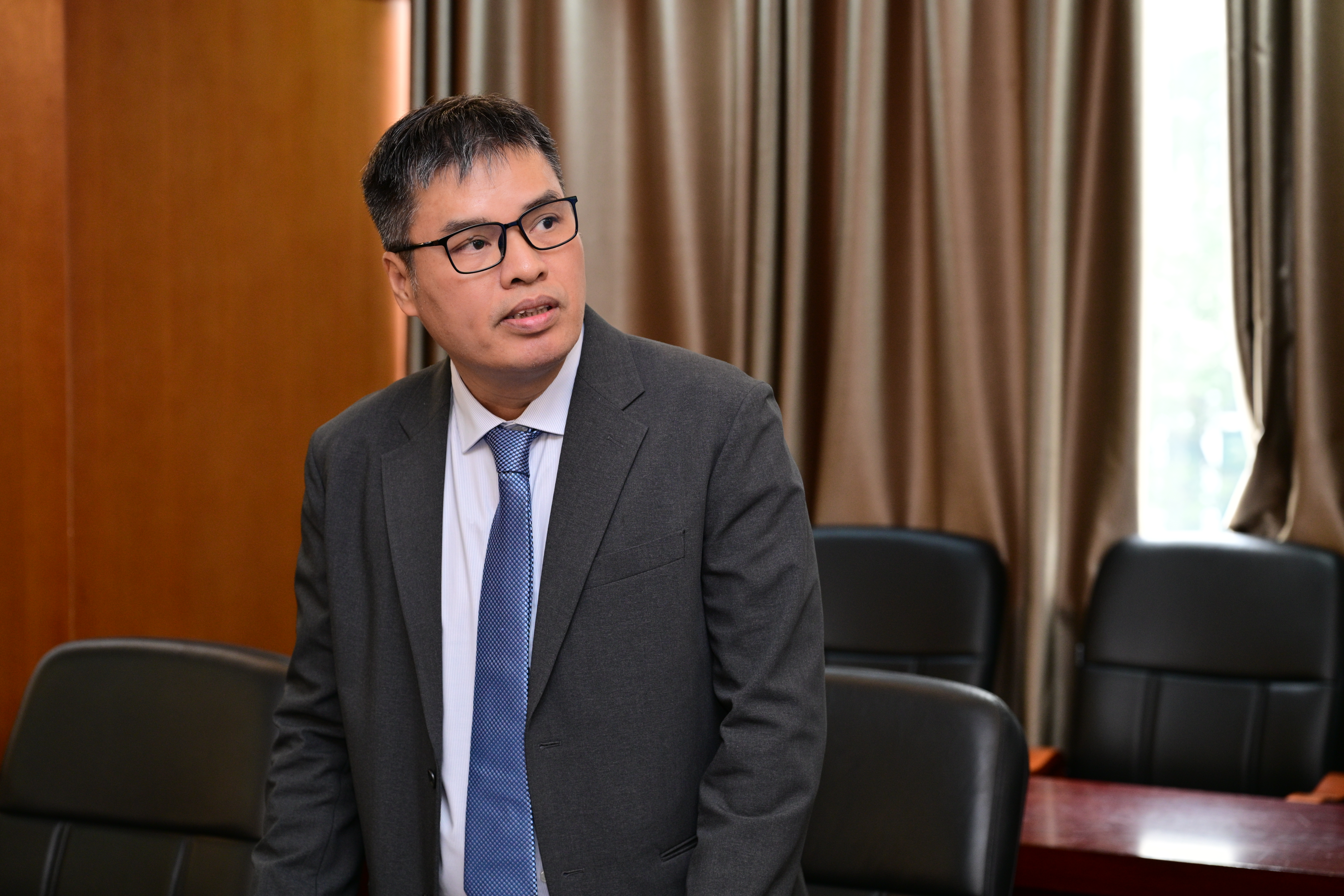
At the defense, the candidate presented the main content and findings of the thesis. The presentation focused on developing deep learning methods for X-ray image classification through image quality improvement, data augmentation, and model design. The committee then discussed and asked questions about the research methods, the reliability of the results, and opportunities for further development.
Using deep learning to analyze and understand X-ray images
X-ray images provide important information for diagnosing and monitoring diseases. However, abnormalities can be difficult to identify because of low contrast or complex image structures. The use of artificial intelligence in X-ray image analysis also faces challenges related to training data and the ability to explain predictions.
To address these challenges, the thesis develops deep learning methods to make better use of information in X-ray images. The approach combines image processing, additional AI training data, and model architectures that analyze both fine details and the overall image context.
The study uses CLAHE, a technique that improves local contrast, to make hard-to-see structures clearer. Generative Adversarial Networks (GANs) create synthetic images for training, while transfer learning uses knowledge from previously trained models. A genetic algorithm helps find suitable hyperparameters, or settings that guide how a model learns.

Scientific contributions and international publications
A key part of the thesis combines two AI models: a Convolutional Neural Network (CNN), which identifies small details in X-ray images, and a Vision Transformer, which analyzes relationships between different image regions. This combination allows AI to consider both details and the overall image, aiming to improve classification accuracy.
The research also uses Grad-CAM to highlight image regions that influence the AI’s predictions. This helps reviewers check whether the AI focuses on relevant areas. However, these visual results must be considered alongside expert assessment to judge whether the predictions are reasonable.
During the research, Pham Ngoc Ha produced five international publications: three papers at Scopus-indexed international conferences in 2023, 2024, and 2026; one article published in 2024 in an ESCI-indexed journal ranked Q3 in Scopus; and one article published in 2026 in an SCIE-indexed journal ranked Q2 in Scopus.
These publications share the thesis findings with the international scientific community and contribute to the development of deep learning methods for medical image analysis.
A scientific foundation for further research
By combining image quality improvement, data augmentation, and model design, the thesis helps address several challenges in AI-based X-ray image classification. It also provides a foundation for further studies on the reliability, explainability, and potential applications of deep learning models in this field.
With appropriate validation using relevant data and real-world conditions, the methods could help doctors classify images more accurately, reduce missed abnormalities, and shorten image review time.
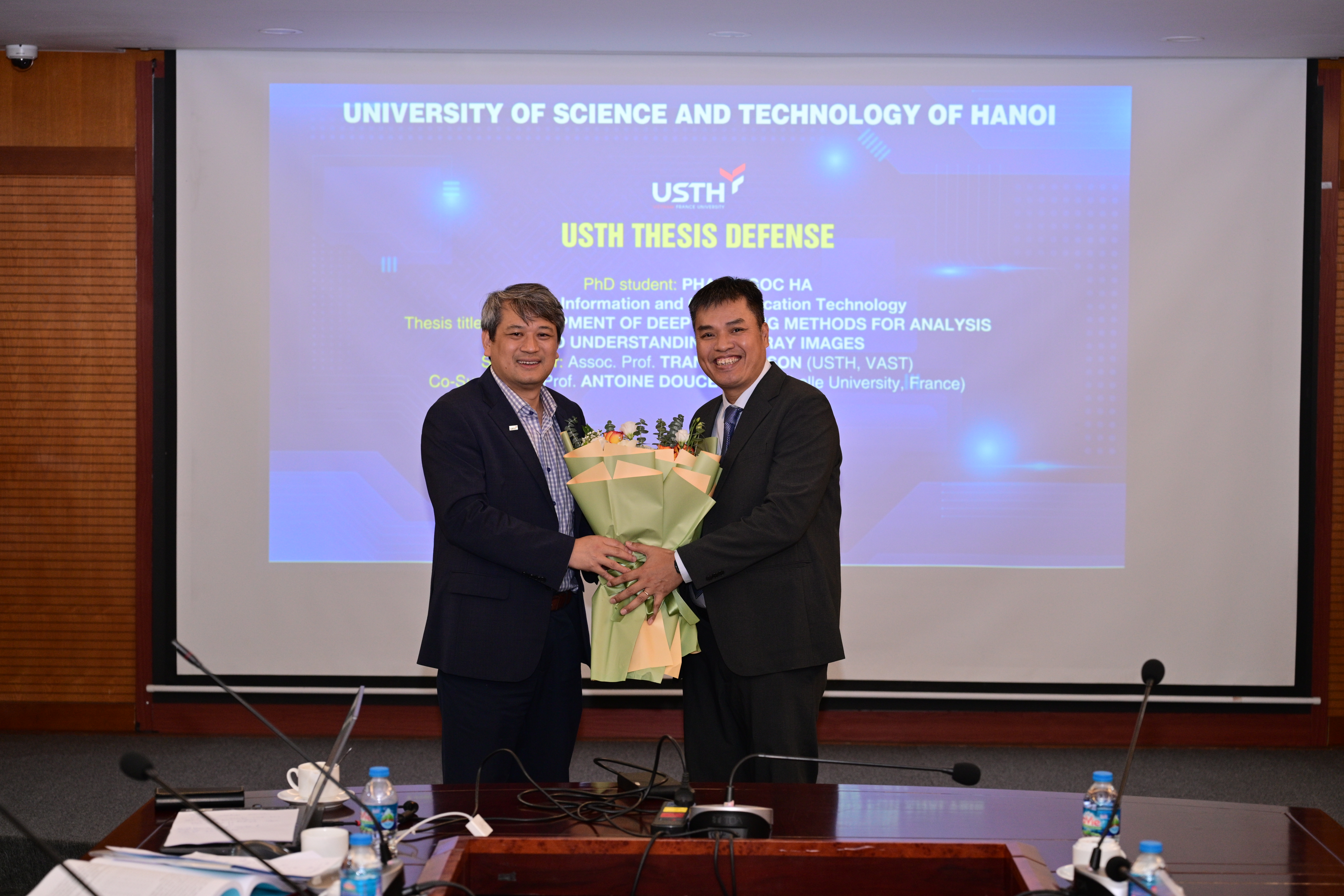
The committee acknowledged Pham Ngoc Ha’s efforts throughout the research and provided feedback to help improve the thesis and guide future work.
USTH wishes Pham Ngoc Ha continued success in research, in further developing these findings, and in contributing to AI applications that benefit the community.